Medical Imaging Software Development
IT Craft brings 20+ years in healthcare IT to build medical imaging software that backs doctors' expertise with sophisticated algorithms.
 Overall review rating
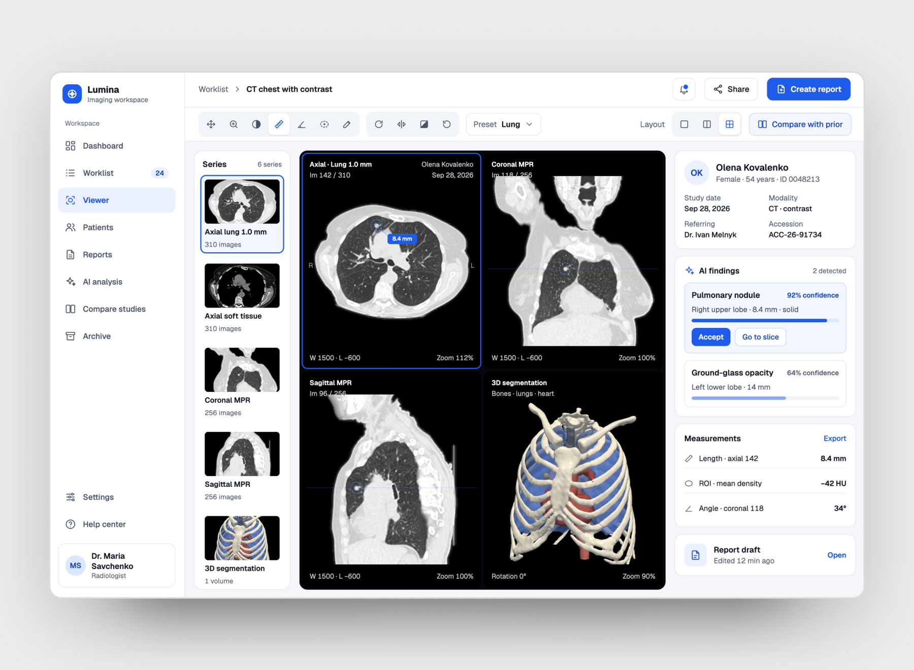
Medical imaging software helps healthcare professionals see a patient’s health clearly, so diagnoses become more accurate and treatments more effective. Paired with imaging hardware (X-ray machines, CT scanners, and ultrasound devices), the software acquires, processes, and analyzes medical images. It enhances image quality, automates abnormality detection, and uses AI for advanced diagnostics.
Cost and Timeline to Develop Medical Imaging Software
The cost to develop medical imaging software usually ranges from $30,000 to $500,000+, with development timelines of 2 to 15+ months. Your final budget and schedule depend on software complexity, the inclusion of AI diagnostic tools, integration requirements, and the target regulatory pathway.
Cost & Timeline Breakdown by Complexity
| Software Complexity Tier | Estimated Cost Range (USD) | Estimated Timeline |
|---|---|---|
| Basic DICOM Viewer | 30,000–80,000 | 2–4 months |
| Single-Modality AI Software | 80,000–200,000 | 6–10 months |
| Enterprise Multi-Modality AI Platform | 150,000–500,000+ | 10–15+ months |
Key Cost Factors
The price of a medical imaging project mostly depends on:
- The complexity of DICOM compliance and integration features.
- The customization level of imaging and visualization tools.
- Security measures for patient data protection.
- Scalability and infrastructure requirements.
- The number of integrations with existing healthcare systems (PACS, RIS, EHR).
- Support for multiple modalities and imaging standards.
- The development team's expertise and location.
Essential Features of Efficient Medical Imaging Software
Below is the functionality our clients request most often when building medical image analysis software, from DICOM viewing to HIPAA-compliant data protection:

DICOM Image Viewing and Manipulation

Multi-Modality Support

Advanced Image Processing Algorithms

Image Annotation and Reporting Tools

Tele-Radiology Capabilities

Mobile Access and Web-Based Viewing

Patient Information Management

Secure Image Storage and Archiving

User Access Control and Audit Trails

Compliance with the Health Insurance Portability and Accountability Act (HIPAA)
Integration with Electronic Health Records (EHR)
Benefits of Medical Imaging Software Development
Our team supports researchers, medical innovators, and medical device manufacturers in preventing, diagnosing, and treating diseases. With both manual and automated (neural network-based) analysis of 3D medical images, providers and patients gain:
- Earlier diagnostics and higher cure and survival rates through machine learning systems.
- Validated diagnoses with the help of neural networks.
- Hidden patterns and valuable insights from research-specific algorithms that advance drug development and the study of complex conditions.
- Precise treatment areas defined by segmentation solutions, which minimize damage to healthy tissue during invasive procedures.
- Immersive training, patient education, and advanced diagnostics through 3D imaging integrated with virtual reality (VR) devices.
Why Choose IT Craft for Medical Imaging Software Development
- Extensive healthcare experience: 20+ years in healthcare software development and 2,250+ projects delivered since 2001.
- Medical imaging expertise: image analysis software development, with work on CT, MRI, PET, SPECT, ultrasound, X-ray, and mammography images.
- DICOM and interoperability: hands-on experience with DICOM, HL7 v2, FHIR R4, ICD-10, SNOMED CT, and LOINC for integration with clinical IT environments.
- Medical software compliance: HIPAA-, GDPR-, and PIPEDA-compliant development aligned with FDA guidance on SaMD and IEC 62304 requirements.
- End-to-end development: architecture, development, modernization, integration, AI implementation, testing, and ongoing support in one team.
- Scalable technical capabilities: 330+ development experts.
Our Approach to Medical Imaging Challenges
You strive to supply clinical stakeholders with medical imaging software that makes diagnostics, treatment, and research evolve. Achieving these goals depends on the reliability and proficiency of your partner, so our approach always includes:
Communication in your language
everyone on our team who contacts you speaks healthcare, so we stay on the same page about the results you expect.
Technical experience that matches the clinical need
our engineers master math algorithms and ML frameworks to build high-performing, cost-effective solutions for image processing.
Compatibility across different imaging equipment and systems
we build interoperability that keeps healthcare workflows seamless.
Data security and privacy regulations
we plan compliance from the start, with encryption, access control, and audit logging.
Standardized sharing of medical images
we implement DICOM to standardize and streamline image exchange between systems.
Scalability
our architects design infrastructure that accommodates growing data volumes without compromising performance.
Advanced features with user-friendliness
we keep a balance between functionality and simplicity, so clinicians reach key tools fast.
Addressing these challenges of medical imaging software development helps the software meet the critical needs of healthcare providers, improving patient care and operational efficiency.
Medical Imaging Modalities We Support
Depending on the type of diagnostics a patient requires, medical staff use different imaging devices, from CT scanners to ultrasound machines. IT Craft develops analysis software for the following modalities:
- CT (computed tomography), such as chest or abdomen scans.
- MRI (magnetic resonance imaging), including brain and cardiac scans.
- PET (positron emission tomography) for assessing cancers, neurological, and cardiovascular diseases.
- SPECT (single-photon emission computed tomography) to diagnose and monitor bone, neurological, and cardiac disorders.
- Ultrasound images for disease prevention, diagnosis, and monitoring across medical specialties.
- X-ray (radiography) scans used in injury care, dentistry, pulmonary care, and other specialties.
- Mammography for breast cancer prevention and monitoring.
- Other imaging modalities.
ML-Powered Medical Image Analysis
Machine learning algorithms process large amounts of medical data and identify complex patterns. In medical image analysis, ML is efficient at:
- Diagnosing diseases
- Segmenting medical images
- Identifying tissue types
- Predicting health outcomes
Body Mapping and 3D Medical Imaging
3D image analysis of particular body parts has its own specifics. Our developers build 3D medical imaging solutions that analyze tissues, bones, muscles, and organs in depth, including the:
- Abdominal system (liver, spleen, kidneys, gallbladder, etc.)
- Genitourinary and endocrine systems (urinary bladder, prostate, reproductive organs, thyroid, etc.)
- Cardiovascular system
- Respiratory system
- Brain and spinal cord
- Mammary glands and other body areas
DICOM Medical Imaging Software Development Process
We follow a structured roadmap to deliver DICOM software that meets clinical needs and healthcare standards.
-
Requirement analysis
- Discovery of healthcare professionals' needs.
- Requirements for image viewing, storage, and sharing.
- Definition of project scope and deliverables.
-
Design and prototyping
- Design of software architecture.
- UX/UI design for radiologists and clinicians.
- Wireframes and prototypes.
-
Development
- Front-end and back-end development.
- Implementation of DICOM standards for medical image processing.
- Compatibility with various imaging devices.
-
Compliance and security
- HIPAA compliance review.
- Encryption, access control, and audit trails.
- Protection of patient data privacy.
-
Integration and testing
- Integration with EHR, PACS, and other healthcare systems.
- Testing for bugs, errors, and compatibility issues.
- Performance validation in real-world scenarios.
-
Deployment and maintenance
- Deployment to the live environment.
- User onboarding and training.
- Updates, bug fixes, and feedback-based improvements.
Technologies We Use for Medical Imaging Software Development
Medical image formats
Our solutions support common imaging standards and specialized extensions, including:
DICOM
Analyze
MINC
NIfTI
ECAT7
Image processing methods

Image quality improvement
At the preprocessing stage, we reduce noise, remove artifacts, compensate for spatial aliasing, and enhance contrast. Clearer images help health specialists confirm the right diagnosis and prepare data for automated image analysis.

Segmentation
This method separates a bone, tissue, vessel, organ, or other structure from its surroundings, for example, isolating brain tissue from the skull and peripheral tissue.

Image fusion and registration
To compare images from different modalities (e.g., CT and PET) or from one modality at different timeframes, our engineers apply fusion and registration methods. They help monitor lesion growth and form change, analyze nonlinear tissue deformations, and support interventional procedures.

Quantification
Through quantification, segmented structures on medical images receive diagnostic information about their size, form, texture, morphology, and dynamics over time.
Contributor
-
Healthcare IT Expert & Consultant